Dec 01, 15 · Thoracic radiography is one of the most commonly employed and useful tools in the diagnostic workup of cats with cardiac disease1, 2 It is used for differentiating cats with respiratory distress associated with cardiac disorders from those with respiratory distress associated with primary respiratory disorders 3 The standard views forThere are many indications for thoracic radiography, both for the diagnosis of intrathoracic disease and as a means of screening to determine the extent of systemic diseases This makes it an essential technique in the clinical investigation of many animals However, despite the frequency with which thoracic radiographs are taken, the thorax remains one of the most challenging areasTant when evaluating the structures of the thorax Several examples would include • kVp at 2 mAs for 15cm dog for analog film (400 speed system) or • 80 kVp at 5 mAs for a 15cm dog for a digital plate radiographic system For any dog measuring 15 cm or greater (measured at the liver or thickest part of the thorax), a grid (81, 110
Dyspnee Chez Un Chat
Radiographie thoracique chat
Radiographie thoracique chat-B Radiographie de thorax La radiographie permet de reconnaitre l'image miliaire par la présence d'un semis d'opacités de 1,5 à 3 mm de diamètre de densité et de répartition variable on distingue 2 types a miliaires pulmonaires typiques Ce sont des micronodules caractérisées parThe normal thorax Some basic rules of thumb can be applied to most dog breeds The two breeds that will break all of the rules will be boxers and bulldogs The widest point of the cardiac silhouette on the lateral view in dogs should be between 25 to 35 intercostal spaces For a cat, on a lateral film, the widest point of the cardiac


Toux Et Hyperthermie Chez Un Chat Quel Est Votre Diagnostic
Thoracic radiography is an essential tool in the investigation of both thoracic and systemic disease Despite the fact that radiography is easy to perform, careful technique is required to ensure that highquality films are obtained Poor technique is a common reason for misdiagnosis The first part of this chapter outlines the methods that should be used to obtain thoracic films andCette radiographie thoracique PA, en position droite, a été prise chez une jeune femme qui a consulté à la suite de douleurs osseuses intenses et de fièvre La taille du contour du péricarde (flèche blanche à deux pointes) est nettement accrue Le rapport coeur thorax (ou rapport cardiothoracique) doit être inférieur à 55 %The normal thorax is well suited to radiographic evaluation because there is marked inherent contrast between the airfilled, fluidfilled, soft tissue, and bony structures that comprise the thoracic viscera and thoracic wall As has been stated before, at least 2 orthogonal views of the thorax are required for complete and accurate interpretation For routine evaluation of the thorax
Radiographie du thorax d'un chat atteint de CMH mentairesChez certains chats, aucune anomalie n'est décelable Pour d'autres, certaines modifications peuvent être entendues (souffle, trouble du rythme, galop cardiaque, ) Elles doivent conduire àTo our present level of understanding and based on some 2,000 CT examinations of the thorax, we have described these pitfalls as they relate to the mediastinum, lung, pleura, and miscellaneous areas In general these pitfalls may be created by 1 Normal anatomic variation 2 The level of the CT slice and partial volume effect 3
Retrouvez les livres de radiologie,imagerie médicale rangés par date de parution Tous sur l' Echodoppler, l'Angiographie, l' IRM, scanner, radiothérapie, tomodensitométrie Tous les ouvrages récents et neufs pour les radiologue et manipulateur radio à des prix promotionnelsEstce que la radiographie peut nous aider à mettre en lumière une lymphadénomégalie au niveau du thorax ?Une radiographie du thorax peut confirmer la présence d'un mégaoesophage, évaluer les poumons en cas de suspicion de pneumonie (si le chat a fait une fausse route) La meilleure technique pour confirmer la suspicion est le dosage des anticorps antirécepteurs à l'acétylcholine


Hernies Diaphragmatiques Chez Le Chat Centre Hospitalier Veterinaire Fregis


Docteur C Est Quoi Cette Tache Blanche Sur Le Coeur De Mon Chat Animages
ObjectiveTo evaluate the severity and extent of lung disease using thoracic computed radiography (CR) compared to contrastenhanced multidetector computed tomography (MDCT) of the thorax in calves with naturally occurring respiratory disease and to evaluate the feasibility and safety of performing contrastenhanced thoracic multidetector MDCT examinations in sedated calvesLa formation plus ou moins importante de liquide qui s'accumule autour du cœurThorax normal radiograph DV, illustration relating to dogs including description, information, related content and more ThreeRiversVeterinaryGroup Felis ISSN Related terms All information is peer reviewed


Adenocarcinome Pulmonaire Chez Une Jeune Chatte Centre Hospitalier Clinique Veterinaire Cordeliers A Meaux 77


Dv Guillaume Gory Wizzvet
Medical devices in the thorax are regularly observed by radiologists when reviewing radiographs and CTs Extrathoracic devices tubing, clamps, syringes, scissors, lying on or under the patient rubber sheets, foam mattresses, clothing, hair braImaging Essentials provides comprehensive information on small animal radiography techniques This article is the second in a 3part series covering cervical, thoracic, and lumbar spine radiography The following anatomic areas have been addressed in previous columns;• Sagittal plane of the thorax parallel to the film and perpendicular to the X ray beam • Center of the Xray beam centered on the heart to avoid distorsion of cardiac silhouette Dog positioning Wrong Correct HW disease in DOGS Pathogenesis • Chronic disease • Damages first at the pulmonary


Radiographie Radioscopie Clinique Veterinaire Des Docteurs Martin Granel Beaufils Jumelle Calvisson
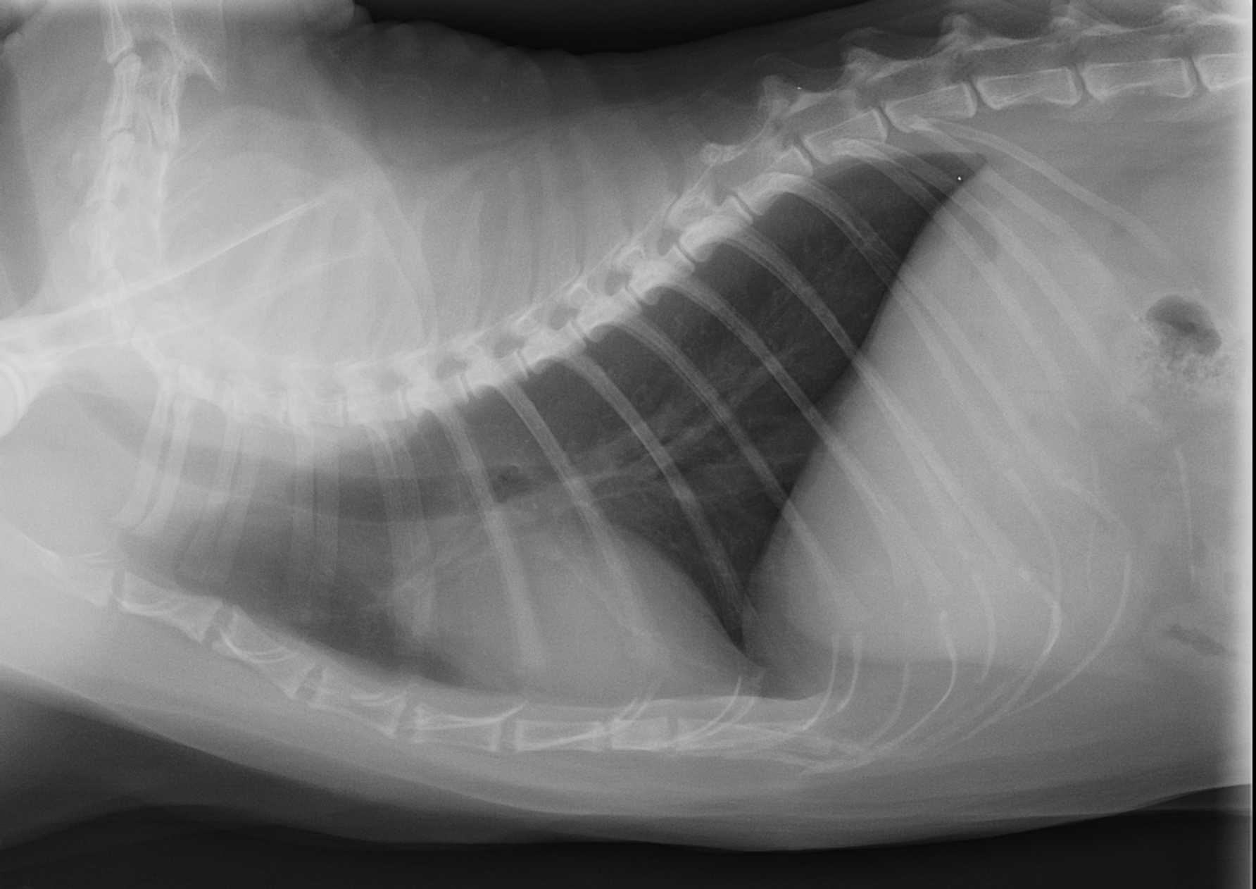


File Radiographie Thoracique Chez Un Chat Jpg Wikimedia Commons
Chez le chien et le chat, les affections du péricarde sont très largement dominées par les péricardites Il s'agit d'une inflammation du péricarde, généralement associée àTo check the degree of thorax rotation, look at the ribs – costochondral junctions should be at the same level and several pairs of the ribs on the centre of the photo should be superimposed on one another The centre of the Xray beam is positioned at the caudal border of the scapula (around the level of the fifth rib) and midway between theGood quality chest films are very important for accurate diagnosis and effective management of the coughing and dyspneic animal Either sign may result from cardiac or respiratory disorders, as well as inflammation, neoplasia, parasitic diseases, trauma,


Anisocorie Chez Un Chat Atteint De Leucose Vision Animale


Veterinaire Nice Imagerie Radiographie Echographie
Explication de contenu d'un radio pulmonaire de F pour les etudiants en medecine et les radiologuesThoracic radiography is one of the most commonly employed diagnostic tools for the clinical evaluation of cats with suspected heart disease and is the standard diagnostic method in the confirmation of cardiogenic pulmonary edema In the past, interpretation of feline radiographs focused on a descripChest radiographs may show hyperlucent areas in the right heart, pulmonary arteries, and systemic veins Signs of pulmonary oligemia, edema, or right heart congestion may also be seen Fat embolism results from trauma to the long bones and pelvis, which can release fat particles and occlude capillaries Production of free fatty acids causes a


Un Cas De Hernie Diaphragmatique Peritoneo Pericardique Chez Un Chien Sciencedirect


Dyspnee Chez Un Chat Europeen Quel Est Votre Diagnostic
RADIOGRAPHIE THORACIQUE NORMALE MariePierre Debray, Nicoletta Pasi, Yaël Amar Hôpital Bichat •Atténuation d'un faisceau de rayons X lors de sa traversée du thorax exposition d'un capteur (film, écran ) •Position du patient TechniqueMême à la radiographie abdominale pour certains d'entre eux (nœuds souslombaires) Mais qu'en estil des nœuds lymphatiques thoraciques?The Xray Anatomy of the Thorax The Xray appearance of the anatomical structures of the thorax in the anterior and posterior views is too well known to require any special description There is, however, one thing about the anteroposterior view which commands some mention


Maladie Bronchique Feline Asthme Pour Ne Pas Le Nommer Animages


Hernie Diaphragmatique Sur Un Chat Vetovie
•La radiographie thoracique = tache difficile • une bonne connaissance anatomique impérative •Buts 02 analyser toutes les structures Eviter les pièges •Atélectasie affaissement, collapsus ou réduction de volume pulmonaire, lobaire ou segmentaire de la radiographie standard du thoraxChat parachutiste Consultée fois Intoxication à la perméthrine chez le chat Une radiographie thoracique permet de mettre en évidence l'air présent dans la cavité thoracique une aiguille est insérée dans le thorax après légère tranquillisation et l'air est aspiré à l'aide d'une seringue Une surveillanceMici et lymphome de bas grade chez le chat Dr Patrick Lecoindre 78 Je sais lire une radiographie du thorax Eymeric Gomes 80 Le top 10 des erreurs à éviter chez vos patients en détresse respiratoire Kevin Le Boedec Boiterie chez le chien trucs et astuces pour arriver au bon diagnostic Guillaume Ragetly 85 L'oeil qui pleure l'antisèche!


Les Differentes Maladies Pulmonaires Vetopedia Conseils Veterinaires


Pneumothorax Et Pneumomediastins Spontanes Chez Un Chat Centre Hospitalier Clinique Veterinaire Cordeliers A Meaux 77
Les erreurs les plus courantes sont l'inclusion d'une partie du thorax seulement (et non du thorax entier) lors du réglage du collimateur, absence de centrage, les prises de vues effectuées lors de l'expiration, le mauvais positionnement (rotation), le mouvement du patient ou des voies respiratoires (flou), la sousexposition ouLa radiographie de thorax ne peut mettre en évidence que les calcifications liées à l'âge, en général bénignes Cette ossification progressive du cartilage costal est régulière au cours de la vie et a même été proposé comme méthode pour donner un âge lors deEffectuer des examens complé
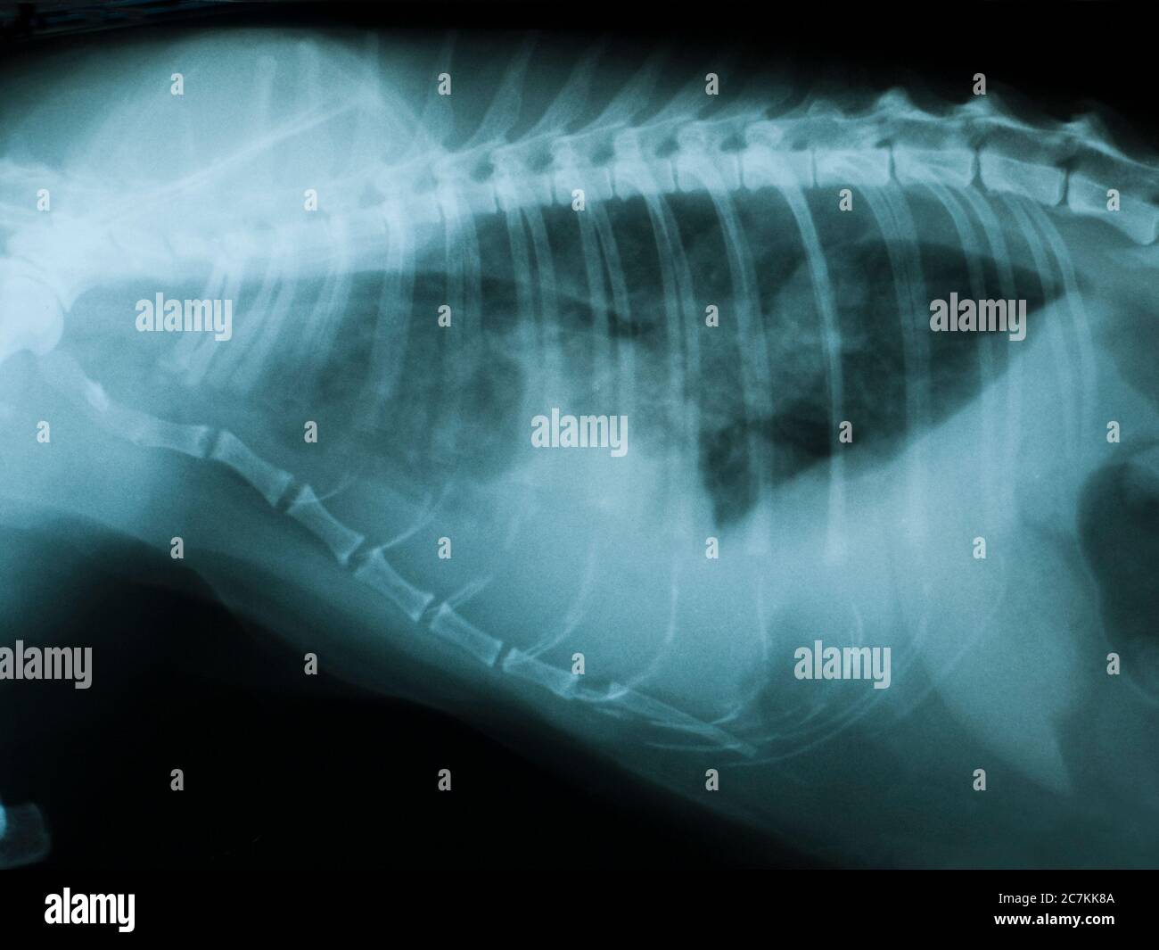


Chat Trachee Banque D Image Et Photos Alamy


Pyothorax Sur Un Chat M Vet
Silhouette in the VD/DV views, the amount of extension was categorized as #25, 25–50, or $50% of the length of the cardiac silhouette, and the side of the extensionwas notedL'asthme du chat évolue généralement sur un mode chronique entrecoupé de crises Le chat asthmatique aura tendance à tousser régulièrement En cas de crise, les symptômes respiratoires deviennent vite alarmants avec un chat qui respire difficilement, gueule ouverte Son thorax se soulève amplement et il peut émettre des sifflementsRadiotherapy tattoos Radiotherapy tattoos or skin markings help make sure external radiotherapy treatment is accurate Tattoo marks During your radiotherapy planning session, your radiographer (sometimes called a radiotherapist) might make between 1 to


Rayons X Des Jambes Du Thorax Et La Tete Avant D Un Chat Normal Image Stock Image Du Humerus Radiologie
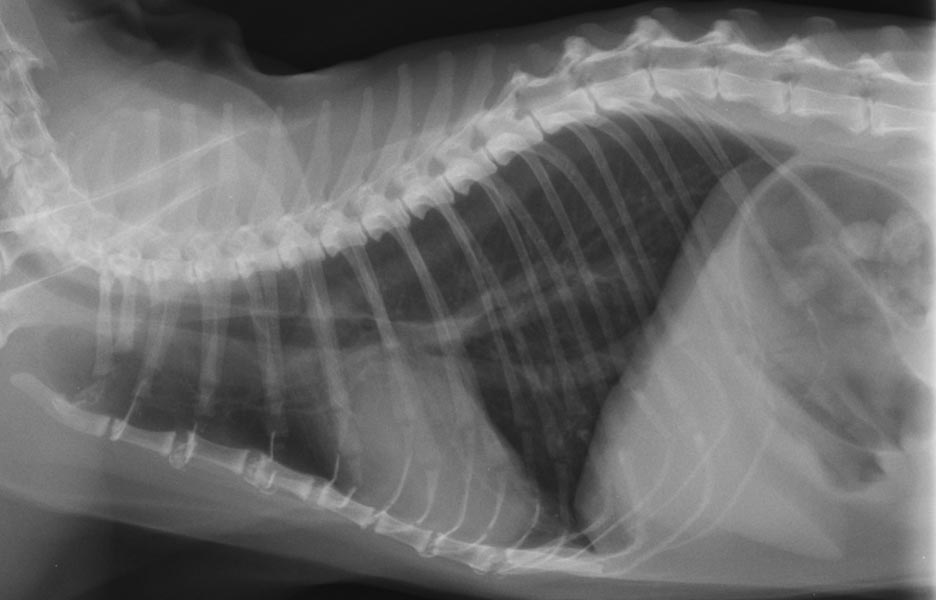


L Asthme Felin Biovet Saint Paul Les Dax
Radio du thorax normale Les objectifs dune bonne lecture dune radio de thoraxDifficultés d intubation du nouveauné pneumo médiastin radio du thorax bronchite Radiographie du thorax de face profil radio du thorax tuberculose Radio publisher to pdf converter free software évident ou petit décollement à bienProfesseur Philippe CHAFFANJONThorax Radiography Chest radiographs (CXR) are routinely abnormal and consistently demonstrate evidence of bronchopulmonary dysplasia2,6,213,218 Common features of chronic lung disease include hyperlucency, hyperinflation, persistent lung hypoplasia, decreased pulmonary vasculature, unknown opacities, persistent mediastinal shifts, and a chronic abnormalA chest CT (computed tomography) scan is an imaging method that uses xrays to create crosssectional pictures of the chest and upper abdomen CT stands for computerized tomography In this procedure, a thin Xray beam is rotated around the area of the body to be visualized Using very complicated mathematical processes called algorithms, the


Suivi De Croissance Osteopathique Qu Est Ce Que La Force De Traction Medullaire Clinique Veterinaire Hermes Plage Et Calypso


10 Signes Precurseurs Du Cancer A Ne Pas Negliger En 21 Homeoanimo
These articles are available at todaysveterinarypracticecom (search "Imaging Essentials") Thorax Scapula,Radiography of the thorax can be problematical due to difficulties eliminating movement blur resulting from breathing High output (high mA capability) Xray machines enable exposure times to be minimized, reducing the risk of movement blur If the machine cannot achieve sufficiently low exposure times, general anesthesia may be requiredIf you like the video than like it, Subscribe it and share with your friends Do not forget to click on Bell icon and never miss the new video If you do not
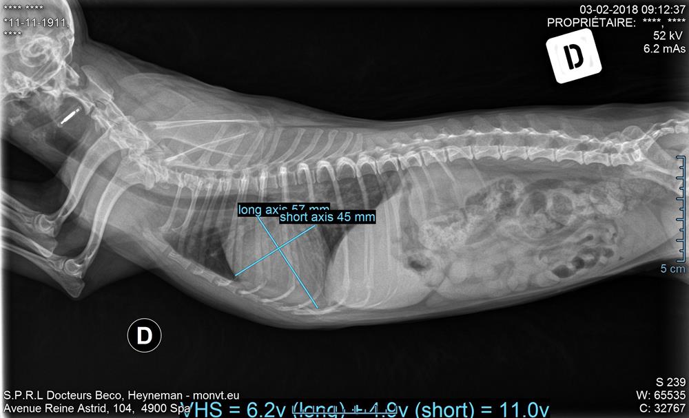


Nouvel Appareil De Radiographie Numerique Pour Une Imagerie De Qualite


Radiographie Du Jeune Chat En Bonne Sante En Vue Laterale Banque D Images Et Photos Libres De Droits Image
Reporting thorax diseases using chest radiographs is often an entrylevel task for radiologist trainees, but it remains a challenging job for learningoriented machine intelligence It's due to the shortage of largescale wellannotated medical image datasets and lack of techniques that can mimic the highlevel reasoning of human radiologistsSi la réponse était non, cet article se révèlerait être des plus ennuyantsThème Radiographie Espèces Animaux de compagnie This book, structured according to the normal anatomical areas in radiological diagnostic imaging (abdomen, neck, thorax, limbs, spine and head), offers both the basis of radiographic interpretation and a


L Oedeme Aigu Du Poumon Oap Sos Veterinaires


Une Blessure Peut En Cacher Une Autre Clinique Veterinaire Hermes Plage Et Calypso
Radiographie du Thorax et de l'Abdomen du Chien et du Chat Body Imaging Thorax and Abdomen reflects the realities of your everyday work it describes the principal anatomic landmarks so that you can orient yourself in the chest and abdomen with speed and confidence, interpret the findings, and make a diagnosisA On the DorsoVentral view NO This is the DorsoVentral view In this case, the accessory lobe region is less aerated because of cranial displacement of the diaphragm secondary by the abdominal organs pressureThe apex of the heart is displaced on the left depending of the aspect of the thorax and the breed of the dogFeb 18, · Ce module de vetAnatomy est un atlas de radioanatomie vétérinaire basé sur des radiographies du chat Les 39 images radiologiques normales de chats ont été réalisées et sélectionnées par Susanne AEB Borofka (PhD dipl ECVDI, Utrecht, PaysBas)


Photo 1 Radiographie Thoracique D Un Chat Atteint D Un Pyothorax Download Scientific Diagram


Anisocorie Chez Un Chat Atteint De Leucose Vision Animale
On which view the accessory lobe region would be best observed ?Radiographie du thorax translation in French English Reverso dictionary, see also 'radiographier',radiophare',radiotélégraphie',radiothérapie', examples, definition, conjugation


Radiologie Clinique Veterinaire Aixiancevet Aix En Provence Puyricard


Un Cas De Pectus Excavatum Traite Chirurgicalement Chez Un Chat Persan De 9 Mois Sciencedirect


Radiographie Imagerie
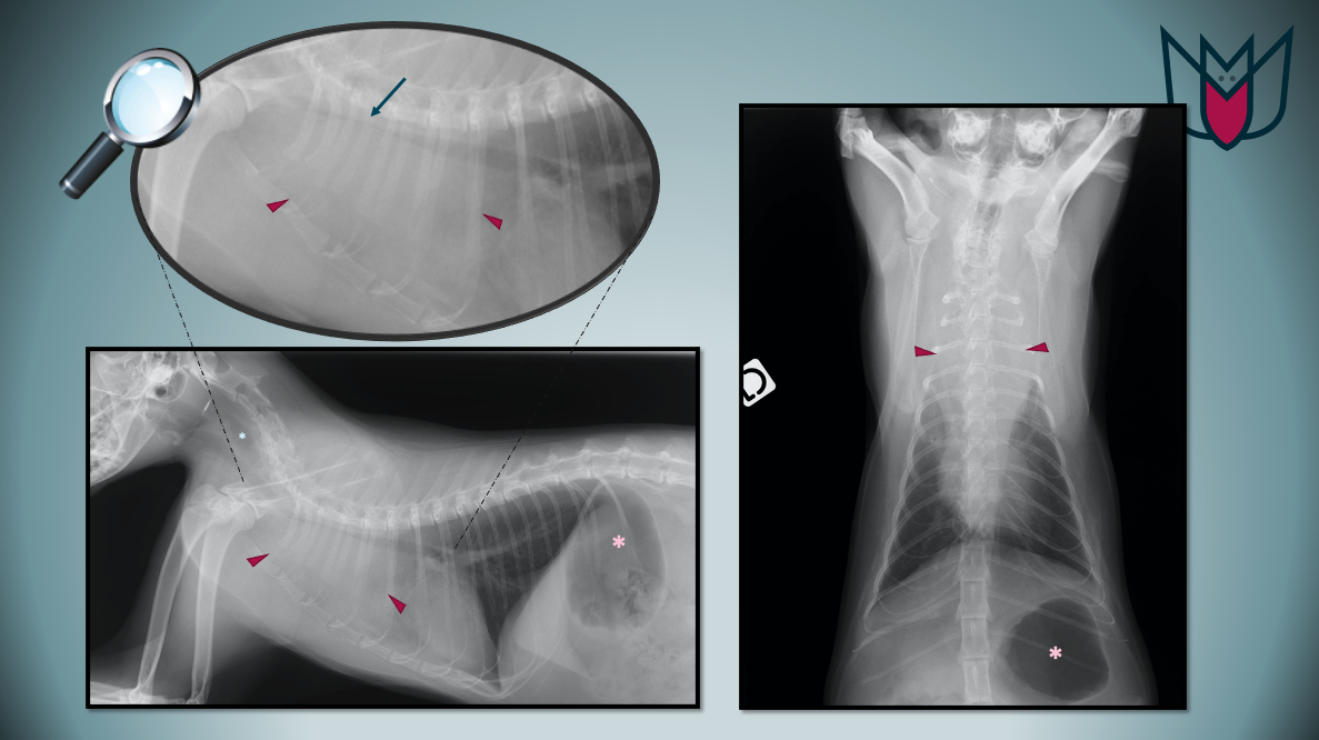


Masse Mediastinale Chez Un Chat


Fiv Felv Case Study Idexx France


Toux Et Hyperthermie Chez Un Chat Quel Est Votre Diagnostic


Imagerie Clinique Veterinaire De Miribel


Pyothorax Chez Un Chat Centre Hospitalier Clinique Veterinaire Cordeliers A Meaux 77


Maladie Bronchique Feline Asthme Pour Ne Pas Le Nommer Animages
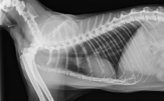


Radiographie Clinique Veterinaire Du Cours A Nantes


Pyothorax Chez Un Chat Centre Hospitalier Clinique Veterinaire Cordeliers A Meaux 77


Radiographies Du Chat


Chylothorax D Origine Idiopathique Chez Un Chat Interet De La Lymphangiographie Scanner Chvsm


Mercredi Imagerie 16 Dirofilariose Chez Un Chien Antillais


Chylothorax Chien Neuilly Sur Seine Paris


Radiographie Thoracique Images Normales Questions Et Images Fournises Par Dr Franck Durieux Dip Ecvdi Aquivet Radiographie Thoracique Images Normales Imagerie Diagnostique Et Outils Sessions Cardio Academy


Pdf Traitement Du Pectus Excavatum Chez Le Chat


Dyspnee Chez Un Chat


Que Peut On Voir Ne Peut On Pas Voir A L Examen Radiographique Clinique Veterinaire Mairie D Issy


Imagerie Medicale Veterinaire
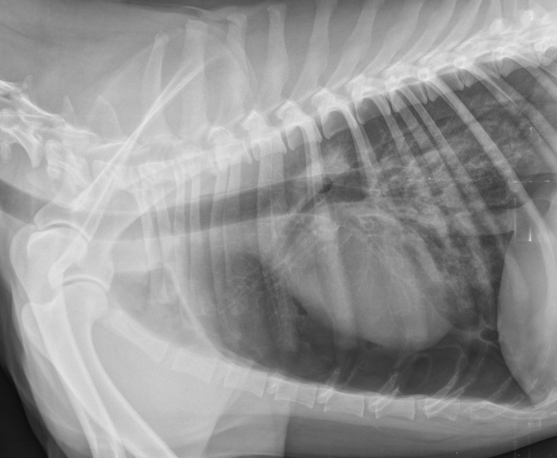


Uc Vet Clinique Veterinaire


Radiographie Radioscopie Clinique Veterinaire Des Docteurs Martin Granel Beaufils Jumelle Calvisson


Radiographies Du Chat


Radiographie De La Cavite Abdominale Et De La Cavite Thoracique Peintures Murales Tableaux Colonne Vertebrale Femur Femoral Myloview Fr


Radiographies Du Chat
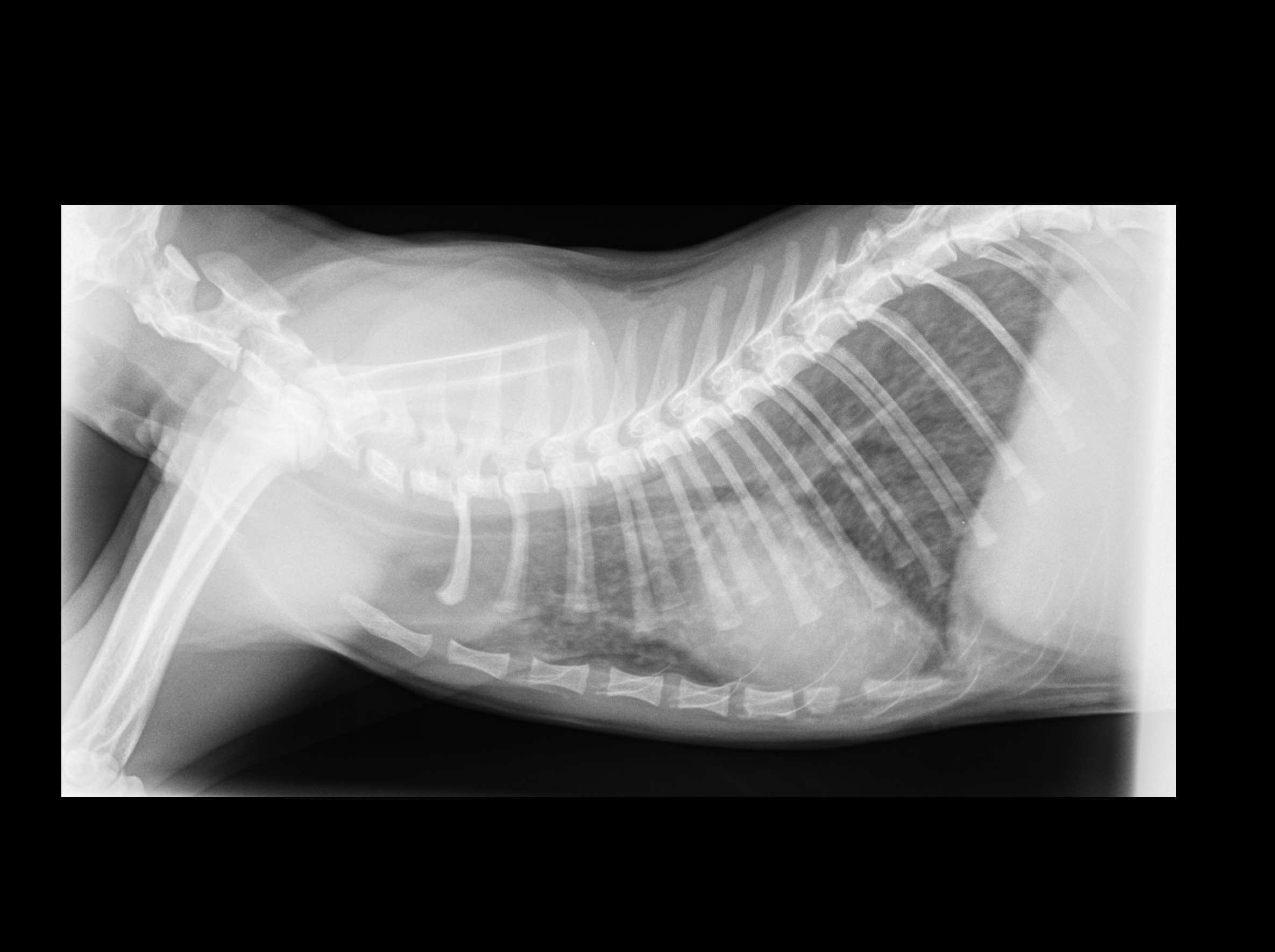


Pneumonie Chat Chaton Et Chat De Race


Pyothorax Chez Un Chat Centre Hospitalier Clinique Veterinaire Cordeliers A Meaux 77


Chat Gonflable La Suite Animages


Radiographie Radioscopie Clinique Veterinaire Des Docteurs Martin Granel Beaufils Jumelle Calvisson


Chylothorax Chez Le Chat Centre Hospitalier Veterinaire Fregis Arcueil


Mercredi Imagerie 18 Hernie Phreno Pericardique Chez Un Chat


œdeme Pulmonaire Cardiogenique Du Chat


Radiographie Radioscopie Clinique Veterinaire Des Docteurs Martin Granel Beaufils Jumelle Calvisson


Chylothorax D Origine Idiopathique Chez Un Chat Interet De La Lymphangiographie Scanner Chvsm


Pyothorax Sur Un Chat M Vet


Faire Une Radiographie Pour Chien Chat Nac Sur Toulon Clinique Veterinaire Pour Nac A Toulon Clinique Veterinaire Du Las
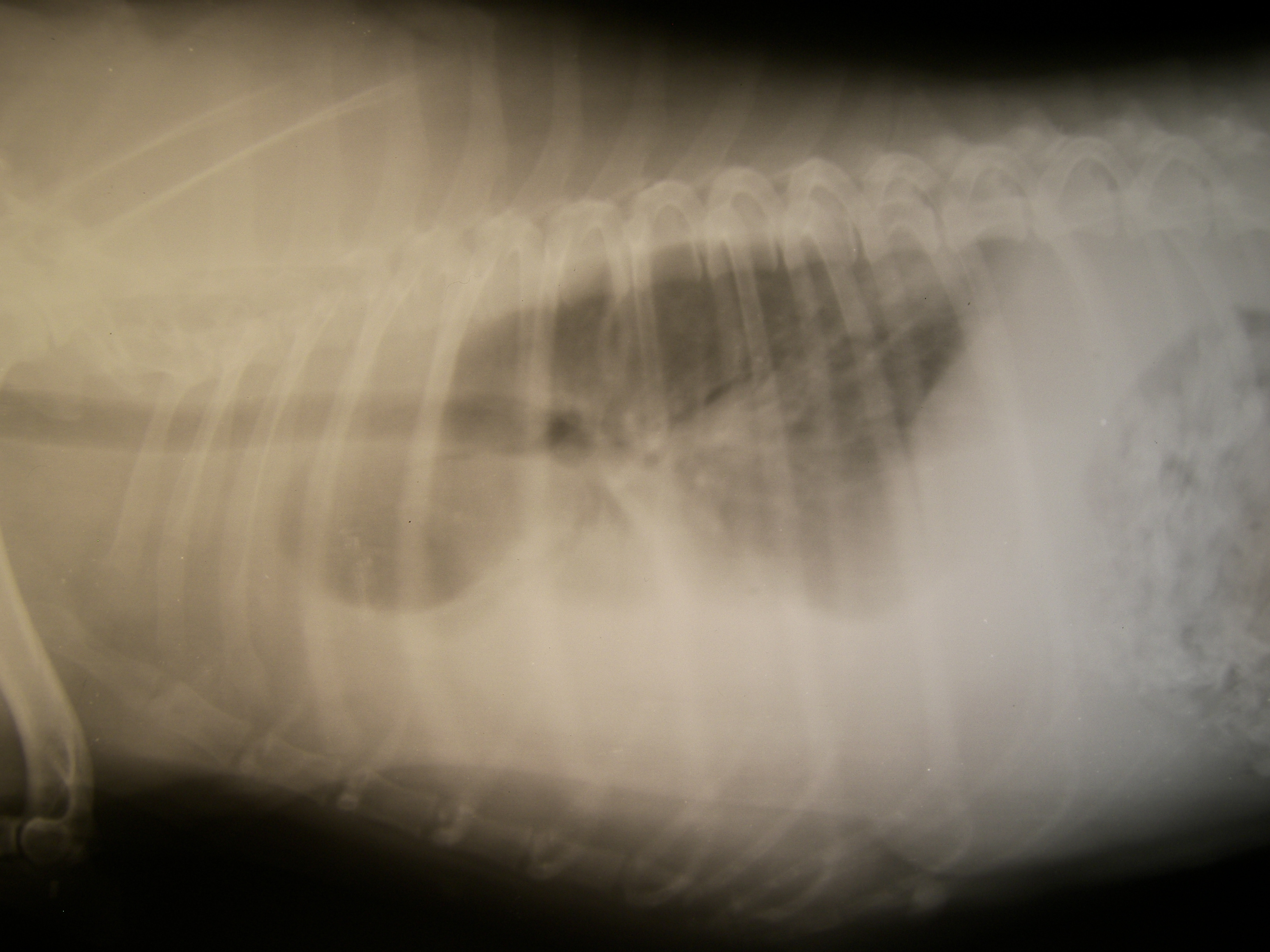


Hemothorax Wikipedia


Radiographie Thoracique Images Normales Questions Et Images Fournises Par Dr Franck Durieux Dip Ecvdi Aquivet Radiographie Thoracique Images Normales Imagerie Diagnostique Et Outils Sessions Cardio Academy


Bon Rayon X Transversal De Thorax De Chat Photo Stock Image Du Nervures Graphique


Dyspnee Chez Un Chat


Faire Une Radiographie Pour Chien Chat Nac Sur Toulon Clinique Veterinaire Pour Nac A Toulon Clinique Veterinaire Du Las


Detresse Respiratoire Chez Les Carnivores


Mon Chat S Est Fait Accidenter Urgences 31
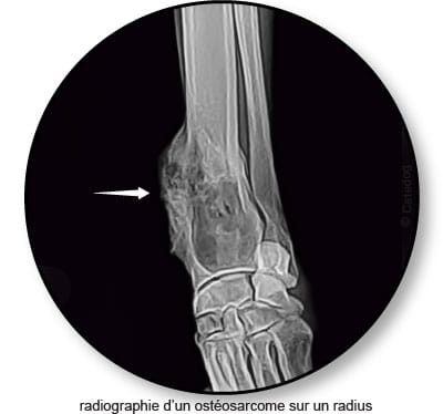


Tumeur De L Os Chez Le Chat Osteosarcome Conseil Veto Illustre Catedog


Radiographie Radioscopie Clinique Veterinaire Des Docteurs Martin Granel Beaufils Jumelle Calvisson


Sos Veterinaire Mon Chat Est Tombe Par La Fenetre


œdeme Pulmonaire Cardiogenique Du Chat


Galerie Illustration De Cas Cliniques Veterinaires Chirurgicaux Medicaux


Diagnostic Et Traitement D Un Pectus Excavatum Chez Un Chat Chvsm


Radiographie Thoracique Images Normales Questions Et Images Fournises Par Dr Franck Durieux Dip Ecvdi Aquivet Radiographie Thoracique Images Normales Imagerie Diagnostique Et Outils Sessions Cardio Academy


La Radiologie Numerique Clinique Veterinaire Des Nympheas


Que Peut On Voir Ne Peut On Pas Voir A L Examen Radiographique Clinique Veterinaire Mairie D Issy
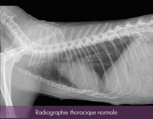


Radiographie Veterinaires Parme


Vetref Clinique De Referesimagerie Interventionnelle Vetref Clinique De Referes


Les Indications De La Radiographie Sont


Radiographie Imagerie


Radiologie Chien Et Chat Vetbookdz
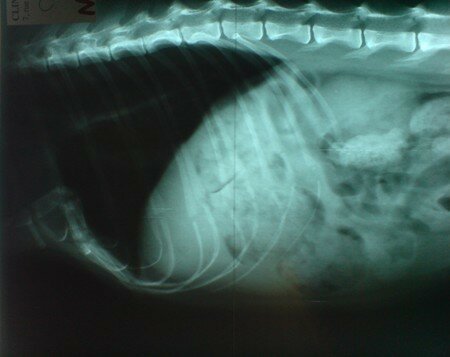


Pectus Excavatum Ou Thorax Creux Reponse A L Article Ci Dessous Elsa Des Nouvelles Et Des Photos 2


Radiographie Imagerie


Contact Vetup Com Auteur A Veterinaire Osteopathie Acupuncture Phytotherapie Homeopathie Veterinaires Physiotherapie Laser Et Imoove Levaillant Chambon 33 Osteopathe Chien Chat Gironde Page 13 Sur 34
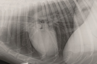


Imagerie Thoracique Du Chien Et Du Chat


Hernie Diaphragmatique Sur Un Chat Vetovie


Diagnostic D Autres Pathologies Des Poumons


Que Peut On Voir Ne Peut On Pas Voir A L Examen Radiographique Clinique Veterinaire Mairie D Issy


Clinique Veterinaire Des Bourgeolles Donzenac Brive Deroulement Radiographie Chien Chat Radiographie Veterinaire


Radiographie Du Thorax Et De L Abdomen Du Chien Et Du Chat Kerstin Von Puckler Florence Le Sueur Almosni Point Veterinaire Livres Medicaux Com


Clinique Veterinaire Du Dr Lustman A Saint Denis La Bronchite Et L Asthme Chez Le Chat Clinique Veterinaire Du Dr Lustman A Saint Denis


Photo 1 Radiographie Thoracique D Un Chat Atteint D Un Pyothorax Download Scientific Diagram


Imagerie Radiologie Et Echographie Clinique Veterinaire Telo Vet


Aucun commentaire:
Publier un commentaire